PATOLOGÍA CEREBELOSA:: MÁS QUE TUMORES Y CAUSAS DEGENERATIVAS. UN ABORDAJE GENERAL.
Resumen
Objetivos Docentes
- Repasar las diferentes patologías que pueden afectar al cerebelo
- Identificar los principales hallazgos de imagen que nos permitan establecer un diagnóstico diferencial entre ellas.
Revisión del tema
El cerebelo es un órgano muy complejo a nivel estructural y funcional. Aunque ocupa la mayor parte del volumen de la fosa posterior, constituye únicamente el 10% de la masa del sistema nervioso central.
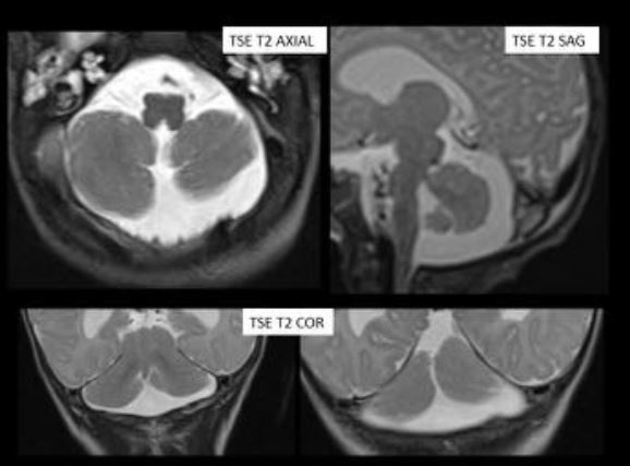
Puede estar afectado por múltiples patologías entre las que se incluyen malformaciones, alteraciones vasculares, tumores, traumatismos, infecciones o enfermedades degenerativas.
En este trabajo haremos un repaso y un diagnóstico diferencial entre las entidades más frecuentes.
Descargas
Referencias
- Rouviere H., Delmas A. Sistema central cerebroespinal en Anatomía humana, descriptiva, topografía y funcional. 9ª ed. Barcelona: Masson; 1987. p. 602-665.
- Bosemani Thangamadhan, Orman Gunes, Boltshauser Eugen et al. Congenital Abnormalities of the Posterior Fossa. Radiographics 2015; 35:200-220.
- Anne G.Osborn and Michael T. Preece. Intracranial Cysts: Radiologic - Pathologic Correlation and Imaging Approach. Radiology: Volume 239: Number 3-June 2006
-Epelman Monica et al. Differential Diagnosis of Intracranial Cystic Lesions at Head: Correlation with CT and MR Imaging. Radiographics 2006; 26: 173-196.
- Kollias, Spyros et al. Cystic Malformations of the Posterior Fossa: Differential Diagnosis Clarified through embryologic Analysis. Radiographics 1993; 13: 1211-1231
- Osborn Anne, Salzman Karen and Barkovich James. Diagnostic imaging. Brain. 2011.
- Bonneville Fabrice et al. Unusual Lesions of the Cerebellopontine Angle: A Segmental Approach. Radiographics 2001; 21: 419 -438.
- Docampo, J et al. Astrocitoma pilocítico. Formas de presentación. Rev Argent Radiol. 2014; 78 (2): 68-81.
- Martínez Leon M.I, Vidal Denis, M and Weil Lara, B. Resonancia magnética en el ependimoma anaplásico infratentorial pediátrico. Radiología. 2012; 54 (1): 59-64.
- Cormier Peter J, Long Eugene R. and Russell Eric J. MR Imaging of Posterior Fossa Infarctions: Vascular Territories and Clinical Correlates. Radiographics 1992; 12:1079-1096.
- Oran 1 et al. Developmental venous anomaly (DVA) with arterial component: a rare cause of intracranial haemorrhage. Neuroradiology. 2009. 51 (1): 25-32
- Arnaout Omar M. et al. Posterior fossa arteriovenous malformations. Neurosurg. Focus 2009: 26 (5): E12