Periné femenino:: revisión de la anatomía y patología
Resumen
Objetivos Docentes
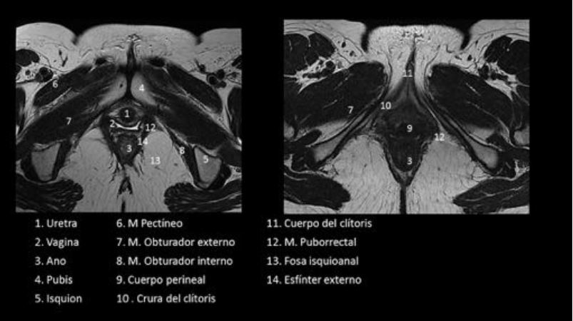
1. Describir la anatomía del periné femenino
2. Conocer las principales lesiones que afectan al periné femenino
3. Revisar el papel de las técnicas de imagen
Revisión del tema
ANATOMIA
El periné es la región anatómica compuesta de partes blandas que cierra la cavidad pelviana, inferior al diafragma pélvico, localizada entre los muslos, la sínfisis del pubis y el cóccix 1,2. Contiene estructuras que soportan las vísceras urológicas, genitales y ano.
Descargas
Referencias
Suh DD,Yang CC, CaoY, Garland PA, Maravilla KR. Magnetic resonance imaging anatomy of the female genitalia in premenopausal and postmenopausal women. J Urol 2003;170:138–144
Hosseinzadeh K, Heller MT, Hoshmand G. Imaging of female perineum in adults. RadioGraphics 2012;32:129-168
Chaudhari VV, Patel MK, Douek M, Raman SS, MD. MR Imaging and US of Female Urethral and Periurethral Disease. RadioGraphics 2010; 30:1857–1874
Berger MB1, Betschart C, Khandwala N, DeLancey JO, Haefner HK.Incidental bartholin gland cysts identified on pelvic magnetic resonance imaging. Obstet Gynecol. 2012;120:798-802.
Viswanathan C, Kirschner K, Troung M, Balachandran a, Devine C, Bhosale P. Multimodality Imaging of Vulvar Cancer: Staging, Therapeutic Response, and Complications. AJR 2013;
:1387–1400
Vulva. In: American Joint Committee in Cancer Staging Manual, 7th ,Edge SB, Byrd DR, Compton CC et al (Eds), Springer, New York 2010.p 379
Pecorelli S. Revised FIGO staging for carcinoma of the vulva, cervix, and endometrium. Int J Gyn- aecol Obstet 2009; 105:103–104
Kochhar R, Plumb AA, Carrington BM, Saunders M. Imaging of Anal Carcinoma. AJR 2012;199:W335–W344
de Miguel Criado J, del Salto LG, Rivas PF, del Hoyo LF, Velasco LG, de las Vacas MI, Marco Sanz AG, Paradela MM, Moreno EF. MR imaging evaluation of perianal fistulas: spectrum of
imaging features Radiographics 2012;32:175-194
García del Salto L, de Miguel Criado J, Aguilera Del Hoyo LF, Gutiérrez Velasco L, Fraga Rivas P, Manzano Paradela M, Díez Pérez de las Vacas MI, Marco Sanz AG, Fraile E. MR
Imaging–based Assessment of the Female Pelvic Floor. RadioGraphics 2014; 34:1417–1439