SÍNDROMES DE COMPRESIÓN VASCULAR ABDOMINOPÉLVICOS (SCVA):: CLAVES DIAGNÓSTICAS
Resumen
Objetivos Docentes
Revisar la fisiopatología, presentación clínica, hallazgos radiológicos y posibilidades terapéuticas de los SCVA, con énfasis en la caracterización anatómica y semiología radiológica.
Revisión del tema
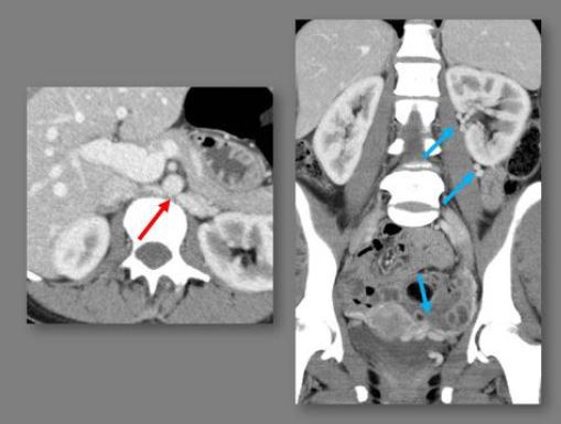
Los vasos abdominopélvicos pueden comprimir estructuras anatómicas adyacentes o ser comprimidos por ellas. Cuando esta condición asocia sintomatología dan lugar al síndromes que incluyen S.ligamento arcuato mediano, S.May-Thurner, S. cascanueces, S.arteria mesentérica superior (S.Wilkie), S.obstrucción de la unión ureteropiélica, y otros síndromes de compresión ureteral.
Descargas
Referencias
Aswani Y, Thakkar H, Anandpara KM. Imaging in median arcuate ligament syndrome. BMJ Case Rep. 2015 Dec 11;2015.
Fong JK, Poh AC, Tan AG, Taneja R. Imaging findings and clinical features of abdominal vascular compression syndromes. AJR Am J Roentgenol. 2014 Jul;203(1):29-36.
Lamba R, Tanner DT, Sekhon S, McGahan JP, Corwin MT, Lall CG. Multidetector CT of vascular compression syndromes in the abdomen and pelvis. Radiographics. 2014 Jan-Feb;34(1):93-115.
Butros SR, Liu R, Oliveira GR, Ganguli S, Kalva S. Venous compression syndromes: clinical features, imaging findings and management. Br J Radiol. 2013 Oct;86(1030):20130284
Wu WL, Tzeng WS, Wu RH, Tsai WL, Chen MC, Lin PC, Tsai IC. Comprehensive MDCT evaluation of patients with suspected May-Thurner syndrome. AJR Am J Roentgenol. 2012 Nov;199(5):W638-45.
Raman SP, Neyman EG, Horton KM, Eckhauser FE, Fishman EK. Superior mesenteric artery syndrome: spectrum of CT findings with multiplanar reconstructions and 3-D imaging. Abdom Imaging. 2012 Dec;37(6):1079-88.
Arthurs OJ, Mehta U, Set PA. Nutcracker and SMA syndromes: What is the normal SMA angle in children? Eur J Radiol. 2012 Aug;81(8):e854-61.
Jeon UB, Chung JW, Jae HJ, Kim HC, Kim SJ, Ha J, Park JH. May-Thurner syndrome complicated by acute iliofemoral vein thrombosis: helical CT venography for evaluation of long-term stent patency and changes in the iliac vein. AJR Am J Roentgenol. 2010 Sep;195(3):751-7.
Moudgill N, Hager E, Gonsalves C, Larson R, Lombardi J, DiMuzio P. May-Thurner syndrome: case report and review of the literature involving modern endovascular therapy. Vascular. 2009 Nov-Dec;17(6):330-5.
Fitoz S1, Ekim M, Ozcakar ZB, Elhan AH, Yalcinkaya F. Nutcracker syndrome in children: the role of upright position examination and superior mesenteric artery angle measurement in the diagnosis. J Ultrasound Med. 2007 May;26(5):573-80.
Braun P, Guilabert JP, Kazmi F. Multidetector computed tomography arteriography in the preoperative assessment of patients with ureteropelvic junction obstruction. Eur J Radiol. 2007 Jan;61(1):170-5.
Park SJ, Lim JW, Cho BS, Yoon TY, Oh JH. Nutcracker syndrome in children with orthostatic proteinuria: diagnosis on the basis of Doppler sonography. J Ultrasound Med. 2002 Jan;21(1):39-45; quiz 46.