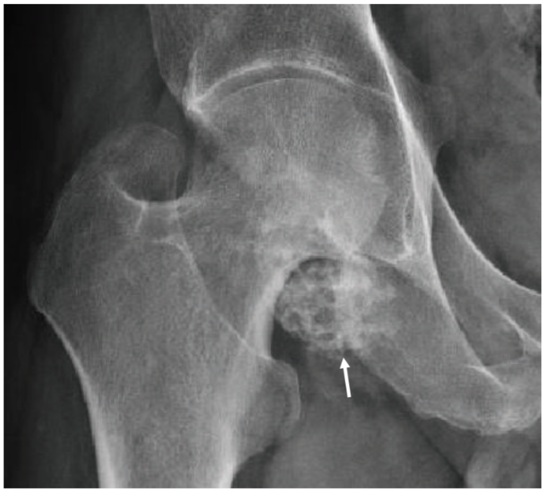
Estudio por imagen de la patología osteo-articular y músculo-tendinosa de la cadera
Resumen
Describir la anatomía radiológica de la cadera, así como los hallazgos por imagen de sus principales causas patológicas, haciendo énfasis en su afectación intra articular.
Descargas
Referencias
Ruiz-Santiago F, Santiago-Chinchilla A, Ansar A, Guzmán-Álvarez L, Castellano-García MM, Martínez-Martínez A, et al. Imaging of hip pain : from radiography to cross-sectional imaging techniques. Radiol Res Pract. Epub 2016.
Llopis E, Higueras V, Vaño M, Altónaga JR. Eur J Radiol. 2012; 81: 3727-36.
Jesse MK, Petersen B, Strickland C, Mei-Dan O. Normal anatomy and imaging of
the hip: emphasis on impingement assessment. Semin Musculoskelet Radiol. 2013; 17: 229-47.
Tibor LM, Sekiya JK. Differential diagnosis of pain around the hip joint. Arthroscopy. 2008; 24: 1407-21.
Schwend RM, Schoenecker P, Richards BS, Flynn JM, Vitale M. Screening the newborn for
developmental dysplasia of the hip. Now what do we do?. Journal of Pediatric Orthopaedics. 2007; 27: 607–10.
Ruiz Santiago F, Castellano García MM, Guzmán Álvarez L, Martínez Montes JL, Ruiz García M, Tristán
Fernández JM. Percutaneous treatment of bone tumors by radiofrequency thermal ablation. Eur J Radiol. 2011; 77: 156–63.